

KDM5A suppresses miR-433 expression
Previous reports have demonstrated KDM5A suppressing downstream genes expression by binding to the promoter of these genes
and demethylating H3K4me3.30 According to the ChIPBase database, KDM5A could bind to the promotor region of miR-433 and
its binding region was in chr14: 100 882 337-100 882 570, of which
its binding site was analysed, e indicating that KDM5A might transcriptionally regulate miR-433 (Figure 3A). RT-qPCR results revealed
that miR-433 level was significantly up-regulated after inhibiting
KDM5A (Figure 3B). To further confirm our hypothesis, we analysed
the expression of miR-433 in HCC tissues and found that the expression of miR-433 was down-regulated in HCC tissues compared with
that of adjacent tissues (Figure 3C) (P <.05). Moreover, ChIP assay
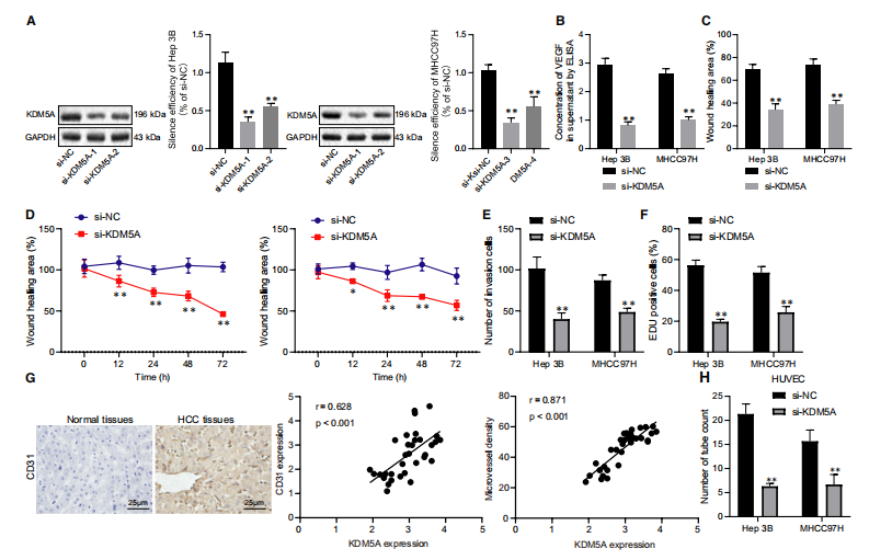
FIGURE 2 Depletion of KDM5A suppressed the proliferative, migrative, invasive and angiogenic capacities of HCC cells. A, silencing
FIGURE 1 KDM5A was significantly up-regulated in HCC tissues and was negatively correlated with overall survival rates. A, the correlation between KDM5A
expression and overall survival rates analysed by GEPIA. B, KDM5A expression
levels in HCC tissues determined by IHC, ×400. C, KDM5A expression in HCC tissues and normal tissues determined by RT-qPCR, N = 110. D, KDM5A expression in Hep3B, MHCC97H and HHL5 determined by RT-qPCR, N = 3. *P < .05; **P < .01, compared to that of normal tissues. Data were shown as the mean ± standard deviation. Statistical comparisons were performed by Tukey's test-corrected one-way ANOVA when more than two groups were compared. The experiment was repeated 3 times
FIGURE 2 Depletion of KDM5A suppressed the proliferative, migrative, invasive and angiogenic capacities of HCC cells. A, silencing
efficiency of independent KDM5A siRNAs in Hep3B and MHCC97H cells determined with RT-qPCR. B, VEGF expression in the supernatant
of Hep3B and MHCC97H cells measured by ELISA. C, effect of KDM5A silencing on the migrative capacities of Hep3B and MHCC97H cells
determined by scratch assay. D, effect of KDM5A silencing on the cell viability of Hep3B and MHCC97H cells determined by MTS. E, effect
of KDM5A silencing on the invasion capacities of Hep3B and MHCC97H cells determined by transwell assay. F, effect of KDM5A silencing
on proliferation determined by EDU assay. G, CD31 expression levels in HCC biopsy specimens determined by IHC. H, the effect of media
from KDM5A silenced Hep3B and MHCC97H cells on the angiogenesis of HUVECs determined by pseudo-tube formation assay. *P < .05;
**P < .01, compared to si-NC. Data were shown as the mean ± standard deviation. Statistical comparisons were performed by Tukey's testcorrected one-way ANOVA when more than two groups were compared. The experiment was repeated 3 times